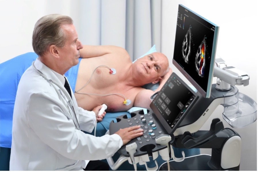
High-End systems are developed to perform at their best in every diagnostic field, which is why every element in them corresponds to the highest technological excellence.
Built with state-of-the-art materials they express the highest diagnostic quality through sophisticated processing algorithms.
High computing power enables the use of very high element density transducers for extremely refined “texturing” of anatomical details.
Next-generation image processing enables 2D, 3D/4D high-resolution ultrasound scans in all fields of application with the highest diagnostic level and a sensational immersive effect.
In addition to quality ultrasound on a traditional basis for the study of organ morphology on a qualitative basis, these systems allow the application of specific softwares dedicated to all body districts to add quantitative and functional information.
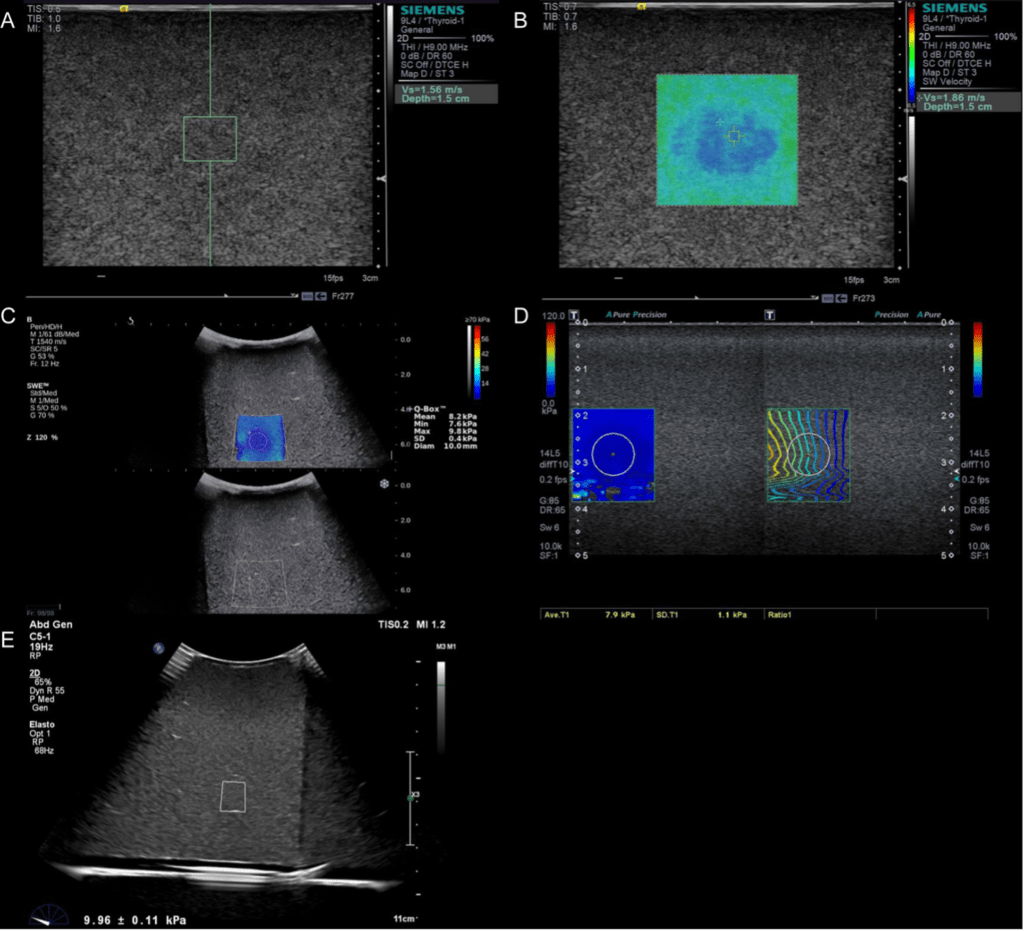
For decades, liver biopsy has been the conventional method for the evaluation of liver disease, more recently thanks to modern innovations elastosonographic function has introduced a comprehensive solution in the evaluation of treatment and monitoring of hepatopathy.
In fact, elastosonography provides real-time 2D assessment of liver parenchyma stiffness with 2D shear-wave (2D-SWE) technology, is easy to perform, noninvasive, and offers the possibility of measurements with reproducible parameters for monitoring steatosis.
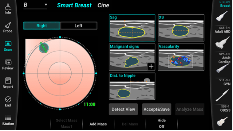
Next-generation ultrasound platforms also assist the operator with artificial intelligence algorithms when studying the breast.
These systems allow for a more complete evaluation of glandular tissue and breast lesions through “lesion detection” functions that enable their characterization of the tissue by automatically determining its morphology and degree of vascularization.
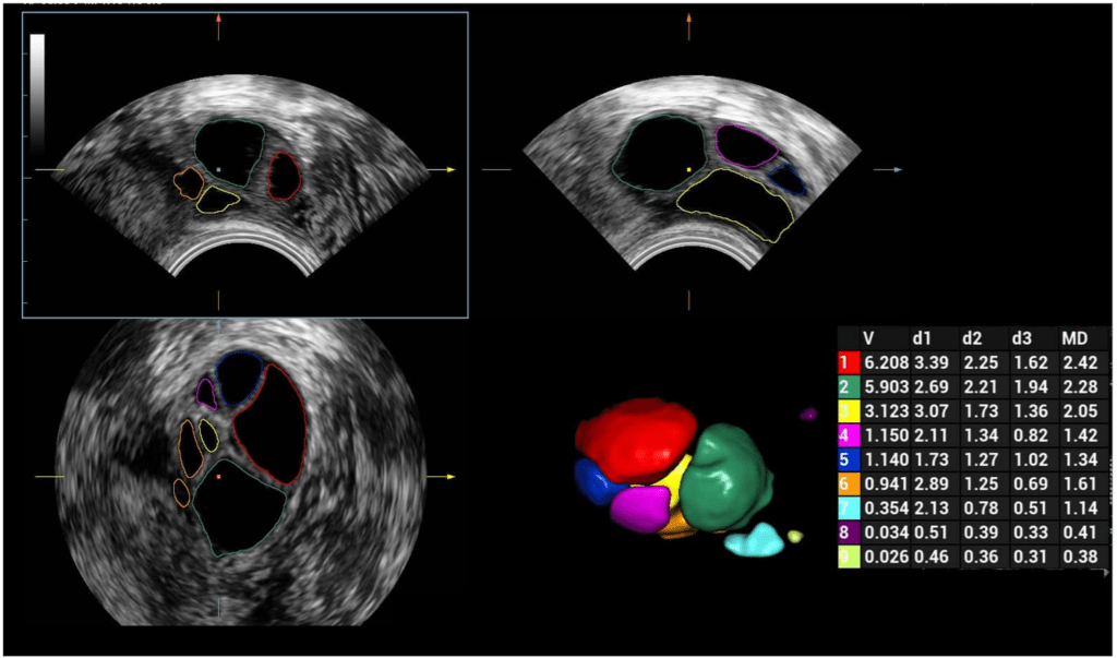
Further technological advancement is achieved through the use of 3D/4D volumetric probes, which can obtain volumetric sectional images on which stratigraphic navigation on the 3 spatial planes X, Y, X is then possible.
This acquisition allows one to obtain a volume consisting no longer of pixels ( two-dimensional ) but of voxcel (3D) that is, obtaining in advantage of the spatial dimension of the structures bringing advantage in the performance of certain ultrasound districts, such as the uterus and ovary.
For example, automated 3D acquisition in follicular monitoring allows follicles to be recognized and classified by volume size improving patient comfort by dramatically reducing examination time.
In this field of application, too, modern technologies offer several diagnostic advantages.
Newly available algorithms contribute to the completion of ultrasound investigations by allowing the operator to collect accurate morphologic as well as hemodynamic information.
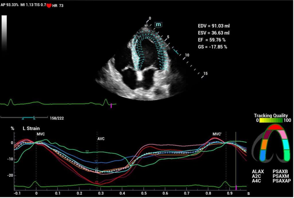
New software in the field of echocardiographic study allows standardization of performance in the evaluation of segmental parietal kinetics with a high predictive degree on organ health status.
We have arranged short-term rental formulas for any type of ultrasound equipment with no commitment to purchase.